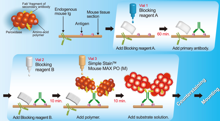
DETECTION SYSTEMS
For Animal tissue
For Mouse
With mouse primary antibodies


| Product Name | Slides | Volume | Code | For use with | Instruction |
|---|---|---|---|---|---|
| N-Histofine® MOUSESTAIN KIT | 50 | 6ml x 3 | 414321F | Mouse primary antibody |  |
| 500 | 17ml x 9 | 414322F |
With rabbit, goat or rat primary antibodies
| Product Name | Slides | Volume | Code | For use with | Instruction |
|---|---|---|---|---|---|
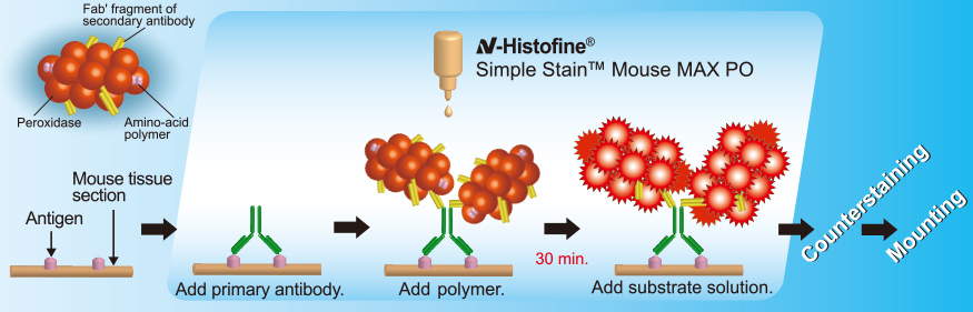
| N-Histofine® Simple Stain™ Mouse MAX PO(R) | 170 | 17ml | 414341F | Rabbit primary antibodies | |
| N-Histofine® Simple Stain™ Mouse MAX PO(G) | 170 | 17ml | 414351F | Rabbit primary antibodies | |
| N-Histofine® Simple Stain™ Mouse MAX PO(Rat) | 170 | 17ml | 414311F | Rabbit primary antibodies | |
For Rat
With mouse, rabbit, or goat primary antibodies
| Product Name | Slides | Volume | Code | For use with | Instruction |
|---|---|---|---|---|---|
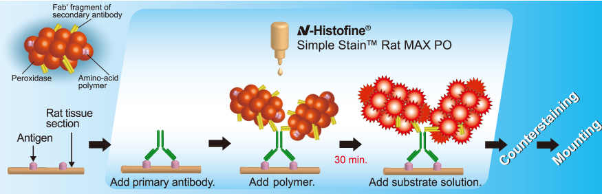
| N-Histofine® Simple Stain™ Rat Max PO(MULTI) | 170 | 17ml | 414191F | Mouse and rabbit primary antibodies | |
| N-Histofine® Simple Stain™ Rat Max PO(M) | 170 | 17ml | 414171F | Mouse primary antibodies | |
| N-Histofine® Simple Stain™ Rat Max PO(R) | 170 | 17ml | 414181F | Rabbit primary antibodies | |
| N-Histofine® Simple Stain™ Rat Max PO(G) | 170 | 17ml | 414331F | Goat primary antibodies | |
Advantage
■ Technical Report
Reference
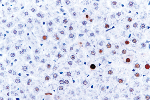
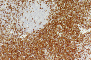
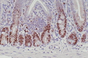
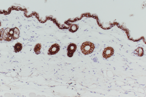
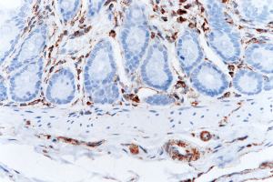
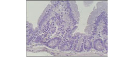
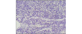
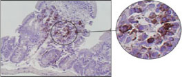
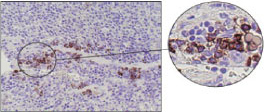
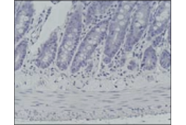
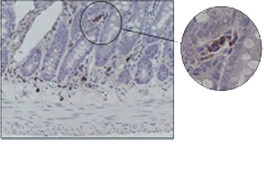
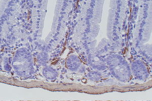
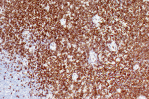
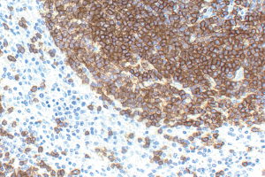
■ Mouse and Rat tissue Sections
- (1) Ezure, K., et al: Glycine Is Used as a Transmitter by Decrementing Expiratory Neurons of the Ventrolateral Medulla in the Rat. The Journal of Neuroscience 23: 8941– 8948, 2003.
- (2) Kitada, M., et al: Translocation of Glomerular p47phox and p67phox by Protein Kinase C-β Activation Is Required for Oxidative Stress in Diabetic Nephropathy. Diabetes 52: 2603–2614, 2003.
- (3) Matsuyoshi, H., et al: Enhanced Priming of Antigen-Specific CTLs In Vivo by Embryonic Stem Cell-Derived Dendritic Cells Expressing Chemokine Along with Antigenic Protein: Application to Antitumor Vaccination. The Journal of Immunology 172: 776-786, 2004.
- (4) Noiri E., et al: Oxidative and nitrosative stress in acute renal ischemia. Am J Physiol Renal Physiol. 281: 948-957, 2001.
- (5) Kazuyo, M., et al: Innate production of TH2 cytokines by adipose tissue-associated c-Kit+Sca-1+ lymphoid cells. NATURE. doi:10.1038/nature08636,2009.
×
×
×