Technical Report2
TOP > Immunohistochemistry > Detection Systems > Technical Report2
Introduction
When the immunohistochemical detection systems for human tissue sections are used for staining on mouse and rat tissue sections, the background staining may be caused due to such reactivity with endogenous immunoglobulins of mouse and rat in the tissue. So that the immunohistochemical detection systems designed for staining on mouse and rat tissue sections were developed. In this report, the background staining is compared between detection systems for mouse, rat and human tissue sections on mouse/rat tissue sections.
Materials & Methods
Materials
– Formalin-fixed paraffin-embedded mouse tissue sections
– Formalin-fixed paraffin-embedded rat tissue sections
–  -Histofine® Simple Stain Mouse MAX PO (Rat) :
a detection reagent designed for staining with a rat primary antibody on mouse tissue sections
– -Histofine® Simple Stain Rat MAX PO (M) :
a detection reagent designed for staining with a mouse primary antibody on rat tissue sections
– -Histofine® Simple Stain MAX PO (M) :
a detection reagent designed for staining with a mouse primary antibody on human tissue sections
Methods
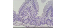
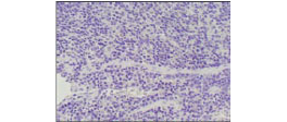
To compare background staining between detection reagents, immunohistochemical staining on mouse tissue sections was performd with -Histofine® Simple Stain Mouse MAX PO (Rat) and -Histofine® Simple Stain MAX PO (M). (Fig.1)
To compare background staining between detection reagents, immunohistochemical staining on rat tissue sections was performed with -Histofine® Simple Stain Rat MAX PO (M) and
-Histofine® Simple Stain MAX PO (M). (Fig.2)
PBS was used instead of a primary antibody in order to identify the background staining caused by detection reagents. DAB solution was used for brown color development.
Steps of immunohistochemical staining
Quenching of endogenous peroxidase
Incubation with PBS for 30 min.
Incubation with detection reagent for 30 min.
Incubation with DAB solution for 10 min.
Counterstaining
Mounting
Results
Fig.1 Mouse tissue sections
N-Histofine®
Simple Stain™ Mouse MAX PO(Rat)
normal colon
normal spleen
N-Histofine®
Simple Stain™ MAX PO(M)
normal colon
normal spleen
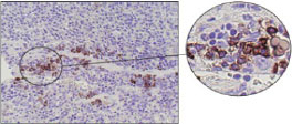
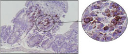
Fig.2 Rat tissue sections
N-Histofine®
Simple Stain™ Rat MAX PO(M)
normal colon
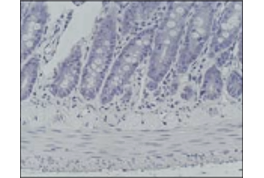
Background staining is NOT observed.
N-Histofine®
Simple Stain™ MAX PO(M)
normal colon
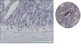
Background staining in plasma cells is observed.
