PRIMARY ANTIBODIES
N-Histofine® ALK Detection kit/ALK Control Slides

| Product Name | Slides | Code | Instruction | Safety Data Sheet |
|---|---|---|---|---|
| N-Histofine® ALK Detection Kit | 20 tests | 417071F |  |
 |
|
Vial NO.1: Blocking Reagent Vial NO.2: Primary Antibody Vial NO.3: Negative Control Vial NO.4: Bridge Reagent Vial NO.5: Peroxidase Labeled Empower Reagent Vial NO.6: Chromogen Substrate Vial NO.7: Substrate Buffer Solution Vial NO.8: Chromogen Reagent Vial NO.9: ALK Antigen Retrieval Solution A Vial NO.10: ALK Antigen Retrieval Solution B |
||||
| N-Histofine® ALK Control Slides | 5 slides | 417081F | |
|
|
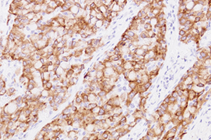
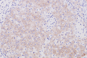
Each slide contains two formalin fixed paraffin embedded cell lines, NCI-H2228 (+: Positive) and SK-BR-3 (-: Negative). |
||||
Reference
ALK Detection Kit
-
(1) Shiota M., et al: Hyperphosphorylation of a novel 80 kDa protein-tyrosine kinase similar to Ltk in a human Ki-1 lymphoma cell line, AMS3.
Oncogene 9: 1567-1574, 1994 - (2) Morris SW., et al: Fusion of a kinase gene, ALK, to a nucleolar protein gene, NPM, in non-Hodgkin’s lymphoma. Science. 263: 1281-1284, 1994
- (3) Takeuchi K., et al: KIF5B-ALK, a novel fusion oncokinase identified by an immunohistochemistry-based diagnostic system for ALK-positive lung cancer. Clin Cancer Res. 15(9): 3143-3149, 2009
-
(4) Takeuchi K., et al: Pulmonary inflammatory myofibroblastic tumor expressing a novel fusion, PPFIBP1-ALK: reappraisal of anti-ALK
immunohistochemistry as a tool for novel ALK-fusion identification. Clin Cancer Res. 17(10): 3341-3348, 2011 - (5) Sugawara E., et al: Identification of Anaplastic Lymphoma Kinase Fusions in Renal Cancer. Cancer, 2012
Sialylated HEG1(SKM9-2)HC
| Product Name | Slides | Code | Instruction | Safety Data Sheet |
|---|---|---|---|---|
| Sialylated HEG1(SKM9-2)HC | 1ml | 498331F | |
|
Reference
HEG1
- (1) Tsuji S, et al. HEG1 is a novel mucin-like membrane protein that serves as a diagnostic and therapeutic target for malignant mesothelioma. Sci Rep. 2017 Mar 31;7:45768.
- (2) Matsuura R, et al. Identification of mesothelioma-specific sialylated epitope recognized with monoclonal antibody SKM9-2 in a mucin-like membrane protein HEG1. Sci Rep. 2018 Sep 24;8(1):14251.
- (3) Hiroshima K, et al. HEG1, BAP1, and MTAP are useful in cytologic diagnosis of malignant mesothelioma with effusion. Diagn Cytopathol. 2020 May 22.
- (4) Naso JR, et al. HEG1 Is a Highly Specific and Sensitive Marker of Epithelioid Malignant Mesothelioma. Am J Surg Pathol. 2020 Aug;44(8):1143-1148.
×
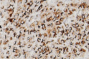
■ Strong Positive Image
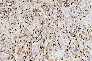
■ Weak Positive Image
×
■ Sialylated HEG1(SKM9-2)

