DETECTION SYSTEMS
For Human tissue
2 step HRP polymer
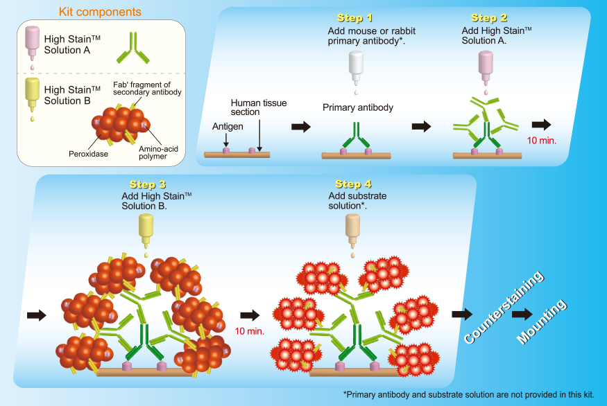
N-Histofine® High Stain™ HRP(MULTI)


| Product Name | Slides | Volume | Code | For use with | Instruction |
|---|---|---|---|---|---|
| N-Histofine® High Stain™ HRP(MULTI) | 170 | 1x17ml each |
414481F | Mouse and rabbit primary antibodies |  |
| 1000 | 6x17ml each |
414483F |
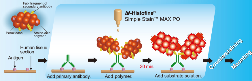
1 step HRP polymer
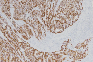
N-Histofine® Simple Stain™ Max PO
| Product Name | Slides | Volume | Code | For use with | Instruction |
|---|---|---|---|---|---|
| N-Histofine® Simple Stain™ MAX PO(MULTI) | 170 | 1x17ml each | 414151F | Mouse and rabbit primary antibodies | |
| 500 | 3x17ml each | 414152F | |||
| 1500 | 9x17ml each | 414154F | |||
| N-Histofine® Simple Stain™ MAX PO(M) | 170 | 1x17ml each | 414131F | Mouse primary antibodies | |
| 500 | 3x17ml each | 414132F | |||
| 1500 | 9x17ml each | 414134F | |||
| N-Histofine® Simple Stain™ MAX PO(R) | 170 | 1x17ml each | 414141F | Rabbit primary antibodies | |
| 500 | 3x17ml each | 414142F | |||
| 1500 | 9x17ml each | 414144F | |||
| N-Histofine® Simple Stain™ MAX PO(G) | 170 | 1x17ml each | 414161F | Goat primary antibodies | |
| 500 | 3x17ml each | 414162F |
| Product Name | Slides | Volume | Code | For use with | Instruction |
|---|---|---|---|---|---|
| N-Histofine® Simple Stain™ MAX PO(MULTI) | 170 | 1x17ml each | 414151F | Mouse and rabbit primary antibodies | |
| 500 | 3x17ml each | 414152F | |||
| 1500 | 9x17ml each | 414154F | |||
| N-Histofine® Simple Stain™ MAX PO(M) | 170 | 1x17ml each | 414131F | Mouse primary antibodies | |
| 500 | 3x17ml each | 414132F | |||
| 1500 | 9x17ml each | 414134F | |||
| N-Histofine® Simple Stain™ MAX PO(R) | 170 | 1x17ml each | 414141F | Rabbit primary antibodies | |
| 500 | 3x17ml each | 414142F | |||
| 1500 | 9x17ml each | 414144F |
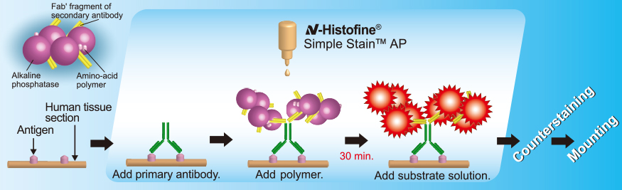
1 step AP polymer
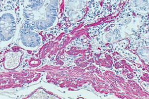
N-Histofine® Simple Stain™ AP
| Product Name | Slides | Volume | Code | For use with | Instruction |
|---|---|---|---|---|---|
| N-Histofine® Simple Stain™ AP(MULTI) | 170 | 1x17ml each | 414261F | Mouse and rabbit primary antibodies | |
| 500 | 3x17ml each | 414262F | |||
| N-Histofine® Simple Stain™ AP(M) | 170 | 1x17ml each | 414241F | Mouse primary antibodies | |
| 500 | 3x17ml each | 414242F | |||
| N-Histofine® Simple Stain™ AP(R) | 170 | 1x17ml each | 414251F | Rabbit primary antibodies | |
| 500 | 3x17ml each | 414252F |
Reference
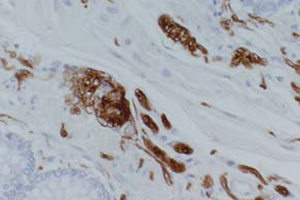
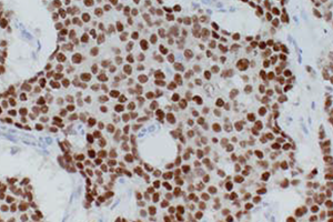
■ Human Tissue Sections
Simple Stain™ MAX PO (MULTI)
- (1) Mokrỳ, et al. Versatility of immunohistochemical reactions: comprehensive survey of detection systems. Acta medica 1996 Apr 39:129-40
-
(2) Yamada K, et al. In vitro assessment of antitumor immune responses using tumor antigen proteins produced by transgenic silkworms. J Mater
Sci Mater Med. 2021 May 17;32(6):58. . - (3) Rahman A, et al. Reduced Claudin-12 Expression Predicts Poor Prognosis in Cervical Cancer. Int J Mol Sci. 2021 Apr 6;22(7):3774.
- (4) Tanaka Y, et al. A Novel Therapeutic Target for Melanoma. Int J Mol Sci. 2021 Jan 19;22(2):976.
- (5) Matsumoto NM, et al. Gene Expression Profile of Isolated Dermal Vascular Endothelial Cells in Keloids. Front Cell Dev Biol. 2020 Jul 29;8:658.
-
(6) Oriuchi N, et al. Possibility of cancer-stem-cell-targeted radioimmunotherapy for acute myelogenous leukemia using 211At-CXCR4
monoclonal antibody. Sci Rep. 2020 Apr 22;10(1):6810. -
(7) Ichinokawa K, et al. Downregulated expression of human leukocyte antigen class I heavy chain is associated with poor prognosis in non-small
-cell lung cancer. Oncol Lett. 2019 Jul;18(1):117-126. -
(8) Yazawa T, et al. Prognostic significance of β2-adrenergic receptor expression in non-small cell lung cancer. Am J Transl Res. 2016 Nov
15;8(11):5059-5070. -
(9) Uchida T, et al. CUL2-mediated clearance of misfolded TDP-43 is paradoxically affected by VHL in oligodendrocytes in ALS. Sci Rep. 2016 Jan
11;6:19118. -
(10) Xing T, et al. Immunity of fungal infections alleviated graft reject in liver transplantation compared with non-fungus recipients. Int J Clin
Exp Pathol. 2015 Mar 1;8(3):2603-14. -
(11) Kaira K, et al. Relationship between CD147 and expression of amino acid transporters (LAT1 and ASCT2) in patients with pancreatic
cancer. Am J Transl Res. 2015 Feb 15;7(2):356-63. -
(12) Toyoda M, et al. Prognostic significance of amino-acid transporter expression (LAT1, ASCT2, and xCT) in surgically resected tongue cancer.
Br J Cancer. 2014 May 13;110(10):2506-13. - (13) Bychkov A, et al. Patterns of FOXE1 expression in papillary thyroid carcinoma by immunohistochemistry. Thyroid. 2013 Jul;23(7):817-28.
Simple Stain™ MAX PO (M)
- (1) Mokrỳ, et al. Versatility of immunohistochemical reactions: comprehensive survey of detection systems. Acta medica 1996 Apr 39:129-40
-
(2) Kaji S, et al. BCAS1-positive immature oligodendrocytes are affected by the α-synuclein-induced pathology of multiple system atrophy.
Acta Neuropathol Commun. 2020 Jul 29;8(1):120. -
(3) Kobayashi S, et al. Image analysis of the nuclear characteristics of emerin protein and the correlation with nuclear grooves and
intranuclear cytoplasmic inclusions in lung adenocarcinoma. Oncol Rep. 2019 Jan;41(1):133-142. -
(4) Kudo I, et al. Particular gene upregulation and p53 heterogeneous expression in TP53-mutated maxillary carcinoma. Oncol Lett. 2017
Oct;14(4):4633-4640. -
(5) Shimura T, et al. MIB-1 labeling index, Ki-67, is an indicator of invasive intraductal papillary mucinous neoplasm. Mol Clin Oncol. 2016
Aug;5(2):317-322. -
(6) Ishiwata T, et al. Enhanced expression of fibroblast growth factor receptor 2 IIIc promotes human pancreatic cancer cell proliferation. Am J
Pathol. 2012 May;180(5):1928-41. -
(7) Tsuji S, et al. Secretion of intelectin-1 from malignant pleural mesothelioma into pleural effusion. Br J Cancer. 2010 Aug
10;103(4):517-23. -
(8) Tanioka Y, et al. Matrix metalloproteinase-7 and matrix metalloproteinase-9 are associated with unfavourable prognosis in superficial
oesophageal cancer. Br J Cancer. 2003 Dec 1;89(11):2116-21.
Simple Stain™ MAX PO (R)
- (1) Mokrỳ, et al. Versatility of immunohistochemical reactions: comprehensive survey of detection systems. Acta medica 1996 Apr 39:129-40.
-
(2) Sudo S, et al. Cisplatin-induced programmed cell death ligand-2 expression is associated with metastasis ability in oral squamous cell
carcinoma. Cancer Sci. 2020 Apr;111(4):1113-1123. -
(3) Kudo I, et al. Particular gene upregulation and p53 heterogeneous expression in TP53-mutated maxillary carcinoma. Oncol Lett. 2017
Oct;14(4):4633-4640. -
(4) Ishiwata T, et al. Enhanced expression of fibroblast growth factor receptor 2 IIIc promotes human pancreatic cancer cell proliferation. Am J
Pathol. 2012 May;180(5):1928-41. -
(5) Tsuji S, et al. Secretion of intelectin-1 from malignant pleural mesothelioma into pleural effusion. Br J Cancer. 2010 Aug
10;103(4):517-23. -
(6) Yokota N, et al. Self-production of tissue factor-coagulation factor VII complex by ovarian cancer cells. Br J Cancer. 2009 Dec
15;101(12):2023-9.
×
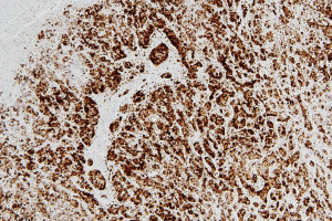
 -Histofine ®
High Stain™ HRP (MULTI)
Competitive Product
Competitive Product
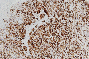
-Histofine ®
High Stain™ HRP (MULTI)
Competitive Product
Competitive Product
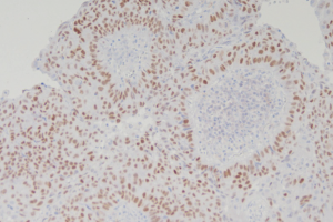
-Histofine ®
High Stain™ HRP (MULTI)
Competitive Product
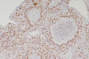
Competitive Product
×
×